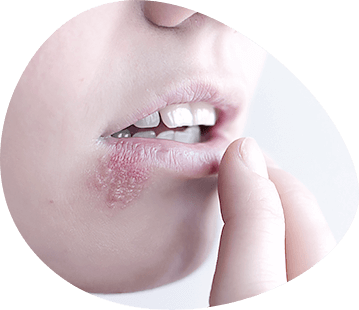
Among the 500 varieties of human pathogenic viruses known to science, herpes simplex virus (HSV) is one of the most common. In recent years, herpetic infection caused by HSV has attracted increasing attention from both the medical and non-medical communities. This is due to the fact that herpes, which has long been known as a rare and unimportant disease, characterized by the rash of painful vesicles on the lips and wings of the nose, has recently significantly increased its importance in human pathology. The increased interest in herpetic infection is also due to a number of other reasons and, above all, the fact that it is now prevalent in various parts of the world, affecting up to 95% of the population, and the virus causing it is able to affect virtually all organs and systems of the human body and cause various forms of infection - acute, latent and chronic recurrent. Clinically, herpes runs as a diverse, complex and often severe disease with the defeat of many organs and tissues, which allows us to consider it as a general systemic disease of the body. In recent years, data on the possible involvement of HSV in the development of some cancers and cardiovascular disease of man began to accumulate.
One of the promising directions is the study of geometric characteristics of cells affected by HSV. The use of traditional projection methods does not give qualitative results, therefore computer image processing is more precise and effective.
Nowadays, computers are widely used in medicine and scientific research, which have become an indispensable attribute of various technical complexes. This also applies to modern control and data acquisition systems, control and laboratory equipment, i.e. any complexes, the main task of which is the processing and interpretation of information coming from the "outside world". The majority of these devices deal with images obtained in one way or another, so the task of image processing and analysis becomes relevant. Methods of processing of images now play a considerable role, especially at working out of systems of an artificial intellect and technical vision.

The image processing process consists of three main steps: image input, processing and analysis. Image input involves improving the signal shape to reduce noise, converting the signal from analog to digital and storing the image. After the input step, the image processing step begins. Image analysis is the identification of objects and obtaining their quantitative and qualitative characteristics.
Herpes and its role in human pathology
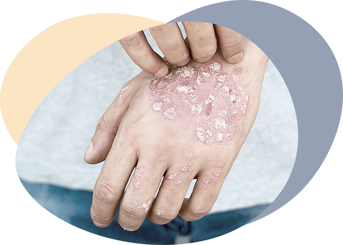
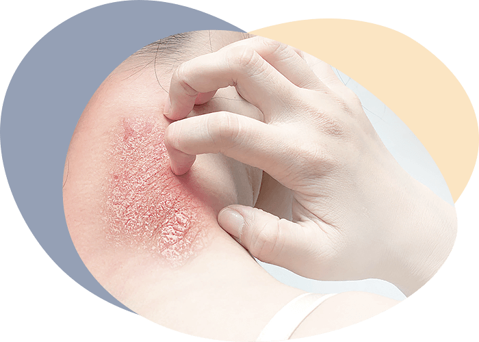
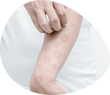
Herpes is one of the leading viral diseases. This is due to the ubiquity of the herpes simplex virus, the variety of clinical manifestations of the disease and, as a rule, its chronic course, as well as the different routes of transmission. Herpes may be accompanied by lesions of the CNS (encephalitis, myelitis, encephalomyelitis), eyes (keratitis, keratoconjunctivitis, uveitis), liver (hepatitis in newborns and adults), mucous membranes (stomatitis, aphthous ulcers, lesions of genitals) and skin (eczema, vesicular rashes). According to the World Health Organization (WHO), diseases caused by HSV rank second (15.8%) after influenza (35.8%) as a cause of death from viral infections It should be noted the already identified etiological and pathogenetic role of HSV (especially HSV type 2 - HSV2) in cervical cancer, which predetermines the cancer aspect of the problem.
On the territory of the former USSR, according to the data collected by virologists and specialists in skin, eye and dental diseases, the number of patients hospitalized in hospitals alone exceeds 2.5 million per year. Significantly more patients are treated in outpatient clinics and polyclinics.
Herpes (from buckwheat, herpes - fever) - the most common viral infection in humans, long existing in the body, mostly in a latent form. It was found that, in addition to the virus affecting humans, it is possible to separate from the organs and materials of sick animals similar in morphology and physicochemical properties viruses that affect only animals.
HSV was first detected in the fluid of herpetic lesions elements in 1912, and the conditions of its passivation on sensitive laboratory animals and cell cultures were later worked out. This made it possible to obtain sufficiently large amounts of the virus to study the structure and properties of HSV, as well as to develop methods for indicating the pathogen to establish the epidemiological features of this infection.
Brief description of the properties of the virus
The HSV virion has a shape close to spherical, and is characterized by pronounced pleomorphism and significant size variations (170-210 nanometers). It contains linear continuous double-stranded DNA (6.5%), protein (70%), phospholipids (20%), carbohydrates (1.6%) and weighs approximately 10-15 grams. The structure of the HSV genome is unusual for DNA-containing viruses. There are 2 types of repetitions in DNA (end and internal) that divide it into two unique covalently linked segments (short and long). The viral genome and associated proteins (nucleoid) are enclosed in an icosahedral protein capsule (capsid) consisting of 162 capsomers, which in turn has an external lipoprotein shell originating from a modified nucleus membrane of the host cell. The lipoprotein shell contains 5-6 viral glycoproteins that are absent in the nuclear and cytoplasmic membranes of uninfected cells. Glycoproteins ensure attachment to the cell and penetration into it. They change the antigenic composition of the infected cell surface, which is important in the pathogenesis of infection and for the induction of the immune response.
According to several authors, virion contains from 24 to 33 structural proteins. Comparative study of proteins and DNA of viral isolates, their biological properties, as well as ways of virus transmission in vivo allowed to distinguish two types of HSV - HSV-1 and HSV-2. Despite the existing differences, representatives of both types of the virus have common group-specific antigen determinants. The presence of antigenic links between HSV strains of the first and second types requires some caution in identifying the viruses released and interpreting the results of sero-epidemiological and diagnostic studies. Moreover, antigenic determinants common to HSV have been identified in all human pathogens of the herpesvirus family: the varicella herpes zoster and Epstein Barra viruses, the cytomegalovirus and the monkey herpesvirus B, which must also be considered in epidemiological studies.
Physico-chemical characteristics
The physical and chemical characteristics of the HSV have been studied quite extensively. The virus is sensitive to drying and heat: it is inactivated at 50-52 0C for 30 minutes, and at 37C - for 10 hours. However, there is evidence that a number of strains of viruses of the first and second types are not fully inactivated even at 56 0C for 1 hour and at 70 ° C for 30 minutes. The study of various factors affecting the thermal stability of the virus has shown that when added to Na2SO4, Na2HPO4, the thermal stability increases, and when added to MgCl2, MgSO4, NaCI, KN2PO4 and KCI it decreases dramatically. The virus, resuspended in aqueous media containing amino acids or protein, was more stable. The greatest thermal stability of the virus was observed at pH 6.5-6.9.
On the other hand, HSV is resistant to low temperatures and maintains biological activity for 1-2 years at -24 0C. The virus is stable at 4°C in 5% glycerin solution. The use of removed milk allows preserving the infectious virus titer without changing it for 5 months at -70°C. Due to the fact that HSV virions consist of lipids by more than 20% (all lipids are localized in the shell), they are easily destroyed by ether, alcohol and other organic solvents (acetone, chloroform, etc.), detergents, proteolytic enzymes (e.g. trypsin). The virus is very resistant to ultrasound and to repeated freezing and thawing. The presence of divalent cations and pH of the medium less than 6.8 increases inactivation of HSV strains when exposed to heat.
Irradiation with ultraviolet and X-rays can destroy the virus even at small doses of exposure. Photodynamic effects of paints have a similar effect. HSV strains tolerate lyophilization well and remain active for 10 years or more in dried form. The virus is well preserved in animal tissues in 50% glycerin solution.
HSV is able to reproduce in almost all known cell lines of vertebrates. When infecting sensitive cell cultures, by intensively multiplying (up to 50 000 - 200 000 virions per cell), the virus inhibits the synthesis of cellular macromolecules, which leads to cell death. In the course of evolution, HSV has developed the ability not only to induce the synthesis of enzymes necessary for its reproduction in an infected cell, but also to coordinate the optimal activity of special enzymatic states of the cell (including phase character) necessary for virus generation, as well as to inhibit and even completely stop the biosynthesis of cell enzymes that are not used for its reproduction. No RNA or DNA polymerase has been found in HSV, but enzymes associated with membranes, such as proteinkinase, have been detected and may be virally specific. HSV DNA has been shown to be infectious.
Morphology and intracellular development
The HSV morphogenesis can be presented as follows. As the virus multiplies, the original viral DNA enters the nucleus; virion DNA is transcribed in the cell nucleus, large viral-specific mRNAs are formed which migrate to the cytoplasm, split into smaller mRNAs associated with ribosomes and form polyribosomes. After synthesis, structural polypeptides are transported to the nucleus and associated with viral DNA, forming a virion capsid. Glycoproteins involved in the formation of the outer shell of virion are embedded in nuclear and cytoplasmic membranes, replacing cell proteins whose synthesis in an infected cell is inhibited. Nucleocapsides, coming out of the cell through specifically modified areas of nuclear and cytoplasmic membranes, capture them as the outer shell of virion HSV. The duration of the HSV reproduction cycle is about 10 hours. Besides its destructive effect on cells in case of acute infection, HSV can have a transformative effect.
The sheathing of the particles has a variety of forms. Sometimes it repeats the hexagonal projection of the capsid. Its diameter varies from 170 to 210 nm. In some cases there are 2 or more nucleocapsides in the common shell. Often the shell is destroyed and many particles are missing. The latter are conditionally called "naked". The particle capsids in the projection have a hexagonal shape with an average diameter of 160 nm. Each facet of it is an equilateral triangle built of 15 subunits of the capsule, arranged at intervals of about 3 nm.
The side of each triangle (icosahedron edge) is made up of 5 capsomers. Each of the 12 peaks is formed by 1 capsomer surrounded by 5 adjacent ones. All other capsomers that make up the edges of triangles are surrounded by 5 neighboring ones. Each capsomer has an elongated prism shape. In cross-section at the tops of the icosahedron they are pentagonal. The other capsomers that make up the surface of the capsid are hexagonal with an inner hole about 4 nm. Thus, the herpes virus capsid is composed of 62 capsomers arranged in 5:3:2 symmetry.
Based on a number of broadly consistent observations, it has been found that the attachment of HSV to cells is quite slow and time is often difficult to measure. The virus is not firmly attached to cells, so about 50% of it can always be separated in different ways.
At the place of adsorption, the cell wall forms a kind of "pocket", which then turns into a vacuum and thus the virus appears in cytoplasm. This is followed by disintegration of the virus, the final result being the release of nucleic acid from the outer shell proteins. Within 10-12 hours after infection, it is not possible to detect any characteristic signs of virus formation in cells. At this time, structural viral proteins and nucleic acids are formed, from which nucleic acid and capsomers are further organized into a single structure, conditionally called nucleocapside, or vironucleon. It is believed that this takes place, apparently, on the principle of self-assembly and is carried out through physical and chemical processes. Apparently, violation of assembly processes leads to the formation of defective forms of the virus.
The development of herpes is accompanied by the formation of intra-nuclear inclusions. These formations are considered by some researchers as a place of virus collection.
Literature data have suggested several possible ways of forming inclusions. For example, viral offspring are known to include no more than 20% of newly synthesized DNA and about 35% of protein. On the basis of these data, it can be assumed that the part of nucleic acid and structural proteins that remains unused in the formation of the virus can form inclusions. HSV causes a gradual inhibition of macromolecular synthesis in the cell. Chromosome damage is evidence of cellular DNA destruction.
Epidemiology of herpetic infection
Herpetic infection is an example of lifelong human carrying of HSV, which can be transmitted during periods of exacerbation by both vertical and horizontal routes.
The spread of herpetic infection not only in Europe, Asia and America, but also among the indigenous populations of territories underdeveloped by Europeans, such as the deep regions of New Guinea, indicate that herpes is an ancient human disease. In natural conditions, only humans are the source of infection. After the primary infection, HSV perpetrates in the body in a latent form for life, and the impact of various factors (hypothermia, insolation, fever, emotional disorders, bacterial and viral infections, etc.) can cause reactivation of latent HSV with the development of relapses of the disease. In this case, the virus is excreted from the ganglia (trigeminal nerve, sacral ganglia, etc.) as a result of a temporary interruption of latency. The length of time during which relapses occur ensures the survival of HSV even in small isolated populations.
The susceptibility of the population to HSV is unusually high. Primary infection usually occurs in the womb, at an early age and most are asymptomatic, leaving behind a strong humoral immunity, which, however, because of the persistence of HSV in the body is non-sterile. Entrance gate HSV - intact or injured skin, lip and mouth mucous membranes, gastrointestinal tract, nose and conjunctivae, genitalia. Both vertical (congenital; transplacental infection) and horizontal transmission by contact, domestic and airborne means is possible. HSV transmission by direct contact implies physical proximity: HSV-1 is most often transmitted by kissing, with saliva, and HSV-2 is mainly transmitted by sexual contact.
Various medical manipulations have a certain epidemiological significance in the spread of herpetic infection. For example, modern complex surgical operations, especially organ and bone marrow transplants, accompanied by massive transfusions of fresh blood, as well as long-term immunosuppressive and radiation therapy often lead to either activation of latent HSV infection or primary infection of patients in contact with viruses or infected materials. Intra-hospital outbreaks of infection are also possible, especially in intensive care wards, premature and newborn departments. Reduced resistance of patients in such wards, constant contact with caregivers and the frequency of various instrumental interventions are factors predisposing to the horizontal spread of herpetic infection.
Pathogenesis and laboratory diagnosis
Primary herpes in almost 80% of patients is asymptomatic. The benign course of the disease is more frequent and ends in clinical recovery. Usually, specific antibodies appear in the serum a few days after the infection begins. More than 85% of children aged 3 years have viral neutralizing antibodies. The presence of antibodies in the serum of people is observed throughout their subsequent lives and does not always protect against secondary or recurrent herpes. Once in the body, HSV is preserved throughout life, periodically causing relapses of disease, which, as in primary herpes, occur with different severity and with different localization of lesions.
In addition to antibodies, the so-called cellular immunity plays a role in the pathogenesis of herpes as a protective factor, in which it is important to adsorb the virus on the cellular elements, suppression of virus reproduction by interferon and other inhibitors, as well as removal of the virus from the body by various means.
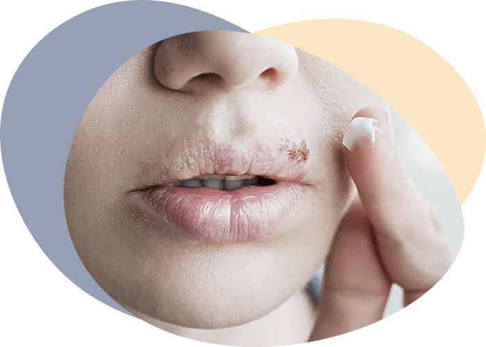
After primary contact of the organism with the virus, the infection is in a "dormant" state or a new exogenous virus infection occurs. The virus, concentrating on the border of the primary infection (most often on the face, in the lip area), again causes a picture of herpetic changes.
HSV remains latent in the human brain after primary infection due to its high neurotropicity. The resting virus can cause encephalitis in humans as a result of activation (e.g. when the temperature increases). Once in the child's body, the virus remains in a latent state for life, and sometimes without relapse it can cause encephalitis.
Numerous experimental and clinical studies on the pathogenesis of herpetic infection indicate that:
- firstly, the leading biological mechanism ensuring the persistence of the virus throughout life in the human body after the first infection and the peculiarities of the course of infection is the HSV latency;
- secondly, significant polymorphism of clinical manifestations of herpes, lesions of many organs and tissues allow considering it as a general systemic disease of the organism.
Clinical polymorphism of herpetic infection is caused not only by the strain features of HSV or the localization of herpetic lesion, but also by the physiological state of the body, its immune response to the penetration and persistence of the virus.
Primary infection
Primary infection is the primary infection of human HSV of any serotype. As already noted, it occurs mostly in early childhood and 80% of patients are asymptomatic. The entry gate to infection is the mucous membranes and skin. The incubation period varies from 1 to 26 days (6-8 days on average). HSV is multiplied at the site of the primary inoculation, where vesicular rashes are formed. The primary virusemia then develops regardless of the mechanism of infection. erythrocytes and leukocytes play an important role in the hematogenic spread of the virus. In the latter, HSV can persist for a long time. Hematogenic disruption of the virus can lead to the development of generalized herpes, especially in individuals with primary or secondary immunodeficiency. HSV concentrations in blood in generalized herpes may reach high levels (10,000,000 infectious units per 1 milliliter of blood). After primary virusemia, HSV actively reproduces in sensitive internal organs and tissues into which it penetrates the capillary barrier, apparently through diapaedesis. The massive release of the virus from the affected organs and tissues into the bloodstream leads to secondary virusemia and acute herpetic infection. The duration of the acute stage usually ranges from 7-14 days. In most cases, individuals who have undergone an acute stage of primary herpetic infection undergo a complete clinical recovery and the body produces specific antibodies, which, however, do not release the body from the virus. In addition to humoral infection, primary infection also stimulates the cellular immune response.
Primary herpetic infection, regardless of the clinical form of manifestation and location of the lesion is accompanied by HSV penetration in the ganglia of dorsal roots and less often - in the vegetative ganglia and ends with their acute infection with subsequent establishment of latency in the neurons.
Latent infection
Latent infection is the presence of HSV in a non-contagious form in any tissue where its replication by specific stimuli can be induced.
The spread of the virus along the nerve pathways plays a crucial role in the formation of latent herpetic infection. Thus, after attachment to the nerve endings, HSV virions are transported within axons at a rate of up to 1.5 millimetres per hour. The mechanisms of acute herpetic infection to latent infection, as well as the form in which HSV is able to exist in this state, are not precisely established. It is known that this transition is caused by a number of factors both from the virus and the host organism, developed during the long evolution of their relationships. Currently, two hypotheses - 'static' and 'dynamic' - have the greatest acceptance. According to the first one, the HSV gene is able to persist under the influence of cell factors (possibly, interferon) in ganglia neurons that do not replicate. According to the second - latent infection is not connected with some special state of HSV, and represents a kind of sluggish process.
In any case, imbalance between the cell and the virus under the influence of provoking factors leads to an increase in replication of HSV, which is expressed in various clinical manifestations - exacerbation of infection. Then a new equilibrium is established between the virus and the cell, as a result of which the formation of HSV stops until some provoking factor again breaks this balanced state. Latent infection is differentiated from acute infection (primary or recurrent) on the basis that in the latter case, HSV can easily be isolated from the tissue samples examined.
Thus, latent infection can be defined as a peculiar form of relationship between virus, cell and organism, in which all known to us protective factors are not able to completely eliminate the pathological process, and HSV, long persevering in the host cells, does not cause their significant destruction.
Returned infection
Along with latency, a characteristic feature of the pathogenesis of herpetic infection is the ability to reactivate latent HSV and develop a relapse (recurrent infection).
Returned infection is characterized by the presence of HSV on body surfaces or in secrets as a result of latent virus reactivation. It has been found that 75% of those who have had primary herpetic infection have relapses throughout their lives, despite a high immune response rate. In relapses of herpetic lesions the resulting local signs and symptoms of the disease are less pronounced, pass faster and, except for certain forms (e.g. herpetic encephalitis, hepatitis, generalized infection), are less frequently accompanied by general clinical manifestations than in primary infection. This is partly due to the fact that the disease develops against the background of the existing specific immunity.
The mechanisms of reactivation of herpetic infection have not been sufficiently studied. Nevertheless, available facts testify that reactivation of latent HSV is to a great extent connected with violations of protective immune mechanisms. Reactivation of latent HSV in persons with primary or secondary immunodeficiency is the most obvious evidence of the leading role of the immune system in the pathogenesis of chronic recurrent herpetic infection. Moreover, in clinical practice it should be considered that herpes is a secondary immunodeficiency disease. There is an opinion that cellular immunity is the leading factor ensuring the prevention of the disease. Its importance in the resistance to HSV is especially clearly seen in congenital defects of the T-system of immunity, thymectomy, immunosuppressant and radiation therapy.
HSV reactivation and recurrence are usually associated with bacterial and viral infections, cooling, fatigue, sunlight exposure, tooth extraction. Thus, for example, according to our data obtained from the analysis of the materials of the Clinic of Eye Diseases of Minsk State Medical Institute from 1978 to 1984, the frequency of relapses of herpetic keratitis increases significantly during influenza epidemics, as well as against the background of an increase in the incidence of acute respiratory viral infections. The reactivating effect of ultraviolet rays on HSV is associated with the release of prostaglandins in the skin. Risk factors include injuries, surgical interventions, changes associated with the menstrual cycle and pregnancy, emotional stress, and others.
The study of interactions between the nervous and immune systems has shown, for example, that the body's defense mechanisms always weaken in a depressed state of morale, and, consequently, conditions are created for a possible reactivation of latent HSV.
Thus, the analysis of the given data testifies to complexity of a pathogenesis of herpetic infection and insufficient studying of leading mechanisms responsible for formation of the latent form of infection and reactivation HSV with development asymptomatic and clinically manifest forms of herpes.
Laboratory diagnostics
HSV is one of the few viruses to detect its etiological role in infectious diseases, using all laboratory diagnostic reactions - from cytological studies to molecular biology methods. The importance of this virus in human pathology and its possible association with certain forms of cancer in humans (HSV), as well as the use of herpes chemotherapy in modern clinical practice, all continue to be important stimuli for the development of new diagnostic techniques.
As with other laboratory tests, those with speed, specificity and sensitivity in detecting HSV have advantages. At the same time, there are many difficulties in interpreting virological test results. This is due to a variety of factors:
- the antigenic, biological variability of HSV isolates;
- different clinical and pathological manifestations of herpes due to the patient's condition (e.g. peculiarities of the flow of herpes in a newborn child or in immunodeficiency conditions);
- subclinical course of the disease in most patients.
The effectiveness of therapeutic and anti-epidemic measures in herpetic infection depends on its timely diagnosis. Clinical differential diagnosis of herpetic lesions of various localizations due to a wide range of manifestations of this infection remains an urgent and important task for practicing doctors of various specialties. Laboratory methods and, first of all, express-diagnostics methods acquire key importance for solving the above mentioned problems.
Depending on the orientation of the search for some or other signs of herpetic infection in the clinical material, these methods can be roughly divided into three groups:
- methods of detection and isolation of infectious HSV;
- methods of detecting viral particles or their components;
- methods of detecting antibodies to HSV.
At the same time, it should be remembered that the quality of the results of diagnostic tests is determined mainly by the time of sampling after the beginning of the disease and largely depends on the technique of collection, compliance with the conditions of primary processing of materials, as well as the interaction of doctors of different specialties (clinicians, virologists, epidemiologists) in the examination of the patient. Thus, the frequency of virus secretion is highest in the case of taking materials for examination in the early stages of the disease and transporting them in the shortest possible time under optimal conditions (at 4°C, in an environment with stabilizing agents - 0.5% gelatin or 0.5% albumin of bull serum). The choice of specific diagnostic test methods is determined by the nature of the course of infection.
Nervous system damage
Neurotropism HSV causes the development of various lesions of the central and peripheral nervous system (CNS and PNS). Complex clinical and virological, immunological and morphological studies have revealed a wide range of manifestations and severity range of herpetic lesions of the nervous system.
Depending on the predominant localization of the pathological process, it is isolated:
- localized CNS lesions;
- localized lesions of the PNS;
- combined lesions of the CNS and PNS;
- combined lesions of the nervous and other body systems.
Depending on the nature of the process course, an acute, subacute and chronic (or recurrent) course is distinguished. In recent years it has been suggested that the subacute and chronic course should be considered a slow form of herpetic CNS infection.
In herpetic infection of the nervous system, the disease may take the form of polyganglioneiritis (cranial, sacral, cranio-sacral), radiculogioneiritis, polyradiculogioneiritis, vegetative polyganglioneiritis, meningitis, encephalitis (meningoencephalitis), myelitis (meningitis), and mental illness. Some researchers have suggested the possible etiological role of HSV in the development of multiple sclerosis, amyotrophic lateral sclerosis, epilepsy, schizophrenia, etc.
Among the most severe manifestations of herpetic infection of the nervous system are lesions of HSV - encephalitis and encephalomyelitis, accompanied by high mortality and disability of surviving patients. In the United States, herpetic encephalitis accounts for 10 to 20% of all CNS viral infections, accounting for 2.3 cases per million population per year. Cases are reported evenly throughout the year. Depending on the age of the patient there are 2 peaks of morbidity: at the age of 5-30 years and over 50 years. It is believed that HSV of the first type is the etiological cause of diseases in 95 per cent of cases.
In the majority of cases herpetic HSV lesions occur in the form of poorly expressed diffuse encephalitis, herpetic meningitis, small or subclinical forms of infection. The course of these diseases is relatively benign and is diagnosed on the basis of virological studies. Over time, such patients may develop chronic nervous diseases and mental disorders. Less often, herpetic lesions take the form of severe encephalitis, among which three forms can be distinguished:
- dysfunctional diffuse meningoencephalitis (the disease is mainly observed in newborns when primary herpetic infection is generalized);
- acute comatose encephalitis in children;
- focal CNS lesions in adults.
Herpetic encephalitis clinic is very similar to other viral encephalitis clinic. However, necrotic encephalitis is characterized by rapid course and adverse outcome. The disease in its first days is considered a respiratory viral infection. Subsequently, the clinical picture develops rapidly. Temperature rises, general cerebral symptoms appear: intense headaches, lethargy, apathy, disorientation in time and space with conscious disturbance, up to comatose state and general irrecoverable convulsions.
Encephalitis caused by HSV have an adverse course and are fatal in 50-90% of cases. Death can occur on day 7-20 of the disease. A case is described where the death occurred one year after the onset of the disease and was due to an acute or recurrent course of the process. Residual events, often in the form of severe CNS lesions, remain in approximately 50% of those who have been ill.
Herpetic encephalitis in children usually occurs with a rapid encephalitis reaction, almost obligatory seizures and consciousness disorders. Menningeal syndrome is relatively weak, only in some cases a moderate inflammatory response is observed. There is no direct correlation between the severity of general infection manifestations and the depth of encephalytic disorders. Temperature normalization does not yet indicate a regression of the infectious process. Early development of comatose state is an unfavorable sign.
Pathogenesis of herpetic encephalitis also differs in different age groups. Thus, in children and young people herpetic encephalitis is the result of primary infection. At the same time, the exogenous virus penetrates into the CNS via nerve fibres from the primary focus (e.g., the olfactor pathway). At the same time, most adults, by the time herpetic encephalitis develops, have clinical or serological signs of herpetic infection with lesions of different localization (e.g., skin or mucous membranes). In these patients, CNS lesions may be the result of reactivation of latent HSV and its subsequent penetration into the CNS via nerve fibres from the primary site of infection, which is directly linked to the CNS for a long time.
The diagnosis of herpetic lesions of CNS and PNS is the most difficult. It is based on a comprehensive examination of the patient in order to isolate HSV from cerebrospinal fluid or brain biopsy and to detect the intracerebral synthesis of antibodies to HSV, especially viral-specific immunoglobulins, or the increase of HSV antibody titer in the disease dynamics.
For early diagnosis of herpetic encephalitis along with specific rapid diagnostic methods it is recommended to use non-invasive para-clinical methods of investigation - electroencephalography and computed tomography.
Articles
Methods of research
Histological preparations
Brain preparations were used in the work (frontal pole field 4). Two main methods of making slices are used to produce these drugs: pouring the brain into paraffin and freezing. Since the brain contains between 70 (in the fibres) and 80% of the water (in the cells), it needs to be dehydrated by replacing the water with the filling material. Compounds with a high affinity for water (e.g. ethanol) can be used to remove it. To minimize tissue damage, these compounds are used in increasing concentrations. However, the brain is reduced in volume by almost 7% during treatment.
Then it goes on to enlightenment and soaking up the tissues. For enlightenment use substances that are mixed with both paraffin and alcohol, such as xylene, in which a piece of tissue is placed by removing it from the alcohol, and the tissue is transferred several times in fresh portions of xylene until all alcohol is replaced by xylene. The fabric is then placed in liquid paraffin, in which it remains until all xylene is replaced by paraffin; this stage is called soaking. Once the block has hardened, the flooded brain is ready to make slices. For a normal examination in a light microscope, the thickness of the slices should not exceed 5-8 µm.
Another method is to prepare frozen slices in a much shorter time than making paraffin slices. To make them, the brain is treated with solutions of formalin with increasing concentrations of sugar. In general, frozen slices are less convenient than paraffin slices because the ice crystals damage the tissue. Frozen fabrics cannot be cut as thinly, and the thickness of the slices prepared from them usually reaches 10 µm.

Since the different components of the living cells have similar optical densities, they all reduce the amplitude of the light waves passing through them to about the same extent, so that none of them seems darker or lighter than the others. Therefore, in order to create sufficient contrast between the different components of cells and tissues to allow them to be examined in an ordinary light microscope, the staining method is used. Dyes are absorbed by different components of cells and tissues to varying degrees and can create contrast in different ways. Usually two dyes are used: first one dye part of the components in one color, and then the other dye the other components in a contrasting color.
Image Analysis System
The main methods of nerve tissue research are removal, stimulation, electrical registration, chemical and histological analysis. For example:
- The location of the nerve structures responsible for a certain behavior can be determined by maximal removal of the areas of the brain where the behavior is maintained and/or minimal removal where it disappears. The functional blockage of nerve centers may serve the same purpose.
- The nerve substrate of the reaction can be analyzed by finding the area and the maximum parameters of electrical and chemical stimulation that cause the same reaction.
- The electrical activity accompanying the behavioural act may reflect the processes important to its realization. Electrophysiological methods can be used to detect the spread of afferent impulses in the brain, the activity preceding the occurrence of an external reaction, or to correlate the probability and/or value of a behavioural and electrical reaction.
- Activation and possible modification of nerve circuits caused by training may be reflected in local changes in mediators, nucleic acids and proteins metabolism.
- Histological description allows qualitatively characterize the structure of tissue and obtain its quantitative characteristics, which allows constructing mathematical models by various functional processes in the studied tissue.
Basics of mathematical image processing
Image analysis plays an important role in histological examination. Geometric measurements of image analysis have emerged from the basic concepts of stereology, so they actively use the concepts and methods of this science. The main geometric concepts are area, perimeter, center of mass, moments of inertia and object orientation. The image in computer memory is discrete and represents a set of pixels, which makes certain corrections to the measurements. The algorithms for calculating area and perimeter are described in the sampling model. According to this model, any flat continuous curve is represented in a discrete image with sampling step h by a set of image points, for which one of the following conditions is met:
- the distance from an image point to the nearest point of the curve intersection with some image line passing through this point is less than 0.5h.
- if this closest distance is 0.5h, i.e. the curve crosses the image line exactly in the middle, it is said that the curve contains an uncertainty point and any of them is included in the discrete representation of the curve.
To avoid laborious viewing of contour points, the peak theorem is used to calculate the area of an arbitrary triangle with all vertices in the orthogonal grid nodes equal to the sum of the number of image points lying inside the polygon and half the number of contour points reduced by one.
An important measurement point is the evaluation or classification of measurement objects. It is necessary to distinguish four general levels of measurement or estimation: nominal, ordinal, interval and relative. The lowest level is the nominal level, where characters such as letters or numbers are used simply to classify objects or phenomena. In this case, the number of measurements falling into different classes under experimental and control conditions is compared using binomial statistics. If it is possible to order the observations so that they are in some relationship, we will deal with an ordinal scale. If, in addition, it is possible to detect intervals between numbers on such a scale, we will deal with an interval scale that has an arbitrary zero point (as in the case of the temperature scale). If the scale also has a true zero point at the beginning, such as a scale of height, mass, then the highest level of measurement, i.e. the ratio scale, will be reached. Parameters measured using a nominal or ordinary scale are processed using non-parametric statistics, while data measured using interval and ratio scales are processed using parametric statistical methods. Population parameters subject to non-parametric statistical procedures do not necessarily have to meet certain conditions, such as normal distribution.
Basic principles of image analysis
The image analysis can be broken down into several main steps:
- image capture and enhancement,
- segmentation (binaryization),
- object detection (identification),
- measurement,
- analysis.
The process of capturing an image is understood as its transformation into an electrical signal suitable for digitization and storage in computer memory as a bit card. The result of digitization is a gray image with quantized space and intensity (gray level).
The process of quantizing space leads to geometric distortions, which are difficult to account for and bring a certain error in measurement. Discretizing the optical density of the object structure in amplitude also leads to various kinds of distortions. Naturally, the quantization of the function is closely related to the quantization of its arguments. Besides, during image capture it is not excluded the occurrence of dot noises, which affect the image quality. For removal of such noises median and averaging filtering is used. Median filtration is a method of non-linear signal processing. A one-dimensional median filter is a sliding window that covers an odd number of image elements. The central element is replaced by the median of all image elements in the window. The median of the discrete sequence a1,a2,...,an for odd n is that element for which there are (n-1)/2 elements larger or equal in size and (n-1)/2 elements smaller or equal in size. Averaging are low-frequency spatial filters, which are most effective for noise smoothing. They are as well as median filters are a sliding window of nn size, where only the central element is replaced.
One of the ways to select the object boundaries is the method of statistical differentiation, where the brightness value of each element is divided into statistical estimation of the standard deviation.
Contrast function is also used to improve the image by varying the gray scale of the image.
By segmentation we mean obtaining a binary image that requires only two brightness levels. There are three different methods of segmentation:
- threshold selection;
- boundary selection;
- regional cultivation.
So, we have a binary image, which has a level corresponding to the empty space, and a level corresponding to objects. Next, we need to select an individual feature for each object. This process is called identification.
As a result of the performed operations we will get an image on which we can make automatic measurements and record the results in the database for further processing.
Image Analyzers
The work of automated image analysis systems is based on methods that relate to the sections of technical vision. The use of such systems provides a number of advantages for the researcher. First, quantitative accounting and classification of features of objects are more objective, as for electronic computers all features are equal. Secondly, calculations and measurements are carried out much faster than manually. Thirdly, during the long work the researcher gets tired and therefore starts to skip the structures he is interested in. In automated analyzers, this is excluded. Fourthly, these analyzers can be used for processing any image, which is their universality.
The BIOSCAN AT image analysis system:
- light microscope;
- television sensor;
- a personal computer with a frame grader;
- digitizer;
- graphic input device mouse.
The computer system for image processing and analysis "Bioscan-AT" developed on the basis of the Minsk State Medical Institute was used in the work. "Biooscan-AT" is a highly integrated flexible analytical system, the main principle of which is to obtain extremely wide opportunities for image processing and analysis due to developed and optimized software with minimum hardware complexity.
The peculiarity of "Bioscan-AT" system is its single-engineering execution. "Bioscan-AT" is a multi-window interactive environment. It contains all functional capabilities of modern image analyzers of well-known western companies at a significant cost reduction. The multi-window graphic environment gives a number of advantages: simultaneous display of several frames, work with large format frames due to scrolling (scrolling) of the image inside the window, saving space on the screen when using overlapping windows. The problem of obtaining the image directly from the camera, which is necessary for finding the object of investigation, adjusting the illumination, pointing at sharpness is solved in the system "Bioscan-AT" at the hardware level by installing a special image input device in the computer. Another important feature of the projected system is a built-in high-level language interpreter that allows you to perform a program of image analysis in automatic mode. The "BioScan-AT" system is equipped with a digitizer - a device for encoding graphic information with increased accuracy, used for system control, image editing and semi-automatic measurements. It is possible to replace the digitizer with the "mouse" manipulator, but this increases the measurement error and eliminates the possibility of working with photos, maps and drawings. Built-in image processing and analysis functions implement most of the currently known approaches to image acquisition, correction, transformation, measurement, reconstruction and storage, including new generation methods of mathematical morphology. The system is in continuous development and is constantly expanding with new functions and capabilities.
The experimental part
Morphological study of herpes simplex virus neuron affection
In the course of the work the analysis of the human cortex affection (field 4) for neurons affection with herpes virus was performed. The material for the study was selected according to the age of 60-70 years, the form of chronic disease, classification of neurons for the first (HSV1) and second type (HSV2) lesions was used. With the help of image analyzer the characteristics of nuclei of all pyramidal neurons were measured and calculated, simultaneously their classification by type of lesion was carried out. The following characteristics were measured: number of cells per unit area, area, shape factor, mean halftone value and nuclear cytoplasmic ratio.
Human brain neurons are a complex object for analysis of their images, it is connected with some problems in preparation of preparations and preparation of tissues. Therefore, a special program in the language of the Bioscan Image Analysis System interpreter has been developed for their selection and classification according to the following algorithm:
- Image capture;
- Adaptive segmentation of the image;
- Removal of false objects, which are not neurons:
- Destruction of objects with the area less than or equal to 30 pixels (noise removal);
- destruction of objects with a shape factor close to 1 (these are glial cells);
- selecting objects with an area greater than or equal to 30 pixels.
- Selection of neurons from a half-tone image by means of conjunctive operation.
- Separation of neuron nuclei by means of threshold segmentation.
- Measurement of nuclei characteristics by type of lesion; parameters for classification are nuclear cytoplasmic ratio, mean value of halftone value.
The measurement resulted in databases describing the geometric characteristics of the affected neurons in each case.
Statistical analysis
This was followed by a basic statistical analysis of the data obtained. Each human organism has many characteristics that distinguish it from others. In this case it is the density of neurons per unit area and the size of the area, which carry the same functional load. Therefore, in order to obtain statistical stability of the studied material characteristics, the percentage of average number of neurons of this class in the unit area was studied. Using average number per unit area allows to get rid of dependence on neuronal density. Using the percentage of average number per unit area allows getting rid of dependence on the cortical area size, which have the same functional load.
In the process of calculations the following values were obtained: mean value, dispersion and standard error.
In order to obtain dependencies of quantitative characteristics of cells with a certain type of lesion on disease activity, regression analysis was carried out. The function allowing to find average (expected) values of one attribute according to the value of one attribute, associated with the first one correlated, is called regression. Statistical analysis of regression is called regression analysis; it allows to judge by how much, on the average, a variable feature can change when changing by a unit of measurement of another, related to it feature.
The relationship between biological traits can be very diverse. In more cases, empirical regressions are expressed by a simple linear dependence equation.
Regression analysis is performed as follows. From several mathematical models, the one that describes the experimental dependence with greater accuracy (by the maximum correlation coefficient, by the minimum standard error, etc.) is chosen. The following models were used to describe the experimental dependencies:
- linear model;
- logistic mode.
The linear model is applicable for dependencies in which the first derivative (rate of change Y) is constant. Logistic model is used to describe limited monotonically increasing and monotonically decreasing experimental dependencies in case of two limits. In this model, the value of parameter "a" defines the lower limit, and the value of parameter "b" defines the distance between the lower and upper limits.
Disease model building
As a result of the analysis of the received data it is possible to draw a conclusion that the process of HSV defeat is not linear, therefore for its characteristic by means of the linear equations it is necessary to use the system which describes both average values of investigated characteristics, and the value characterizing a spread, i.e. dispersion.
It should be noted that the dispersion of the investigated characteristic on each type of the defeated cells is determined by the duration of the disease. At duration of 5 months the dispersion makes approximately 4,88-10-2 (at average value of characteristic 0,5), after 10 months it aspires to 0. I.e. the system has small errors under condition of long duration of the disease (weak activity of the virus), at reduction of duration the system loses its stability.
This leads to the conclusion that the disease activity is characterized by a statistical variation in the number of affected cells, therefore, the system is non-linear. Depending on age, the dispersion changes.
Conclusion
In the course of the work, the lesion of the central nervous system by the herpes simplex virus was investigated. Various geometric characteristics of affected and healthy neurons were measured. Neurons were counted and classified into three classes: healthy, affected HSV1 and affected HSV2. Classification was carried out by morphological features, which are described by geometrical parameters.
In order to get rid of the influence of such characteristics as the sample size and the tissue area under study, a statistically stable parameter in this respect was chosen: the percentage of average number per unit area.
In the course of the study, dependencies were constructed from which it is visible that the system of virus infection is clearly nonlinear. Due to the lack of data to build a nonlinear model, the system was described by equations describing changes in mean value and dispersion.
The equations obtained are fully adequate to the experimental data. This allows us to conclude that the average number of cells affected by HSV1 and HSV2, contrary to popular belief, poorly characterizes the activity of the disease and cannot be a reliable parameter for diagnosis.
The developed model shows a picture of the disease course and will be used in pathology in case of conclusions about correctness and timeliness of the diagnoses, which will allow correcting treatment methods in future.
Obviously, the system of nervous system virus affection is nonlinear in nature, which reverses the existing opinion about the simple picture of the disease and indicates the direction for new scientific research.
By: Dr. Samer Jaber